Advanced Technology

At Drewyer Dentistry, we believe better technology leads to better outcomes for our patients. We have invested in advanced tools that allow our team to diagnose issues earlier, plan treatment with greater precision, and keep patients informed and comfortable throughout every visit. From digital impressions that replace messy trays to AI-supported diagnostics and a fully digital workflow for complex implant cases, our technology allows us to work more efficiently while delivering a higher standard of care.
We are committed to staying current with tools that genuinely improve the patient experience, not simply adopting new equipment for its own sake. Every technology in our office is chosen because it helps us catch problems sooner, plan treatment more accurately, and make each visit more comfortable for our patients in Burtonsville, MD and the surrounding community.
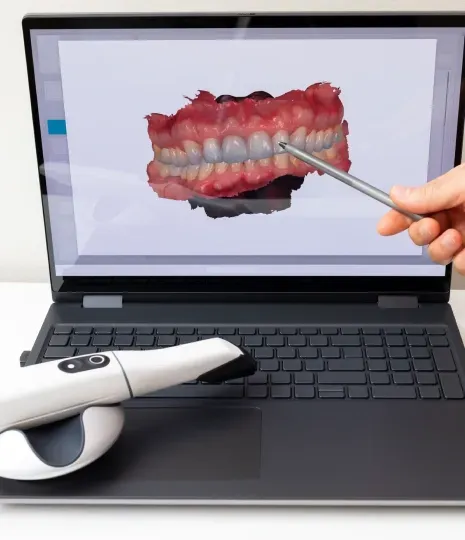
Our intra oral scanners capture precise, digital impressions of your teeth without the thick, uncomfortable putty trays used in traditional impressions. These digital scans are used for everything from crowns and aligners to implant planning, offering greater accuracy and a far more comfortable experience.
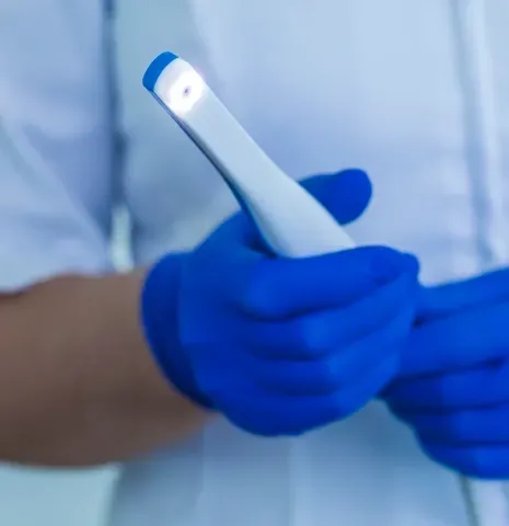
Intra oral cameras are small, handheld devices that let our team capture close-up images inside your mouth in real time. These images appear on a chairside monitor so you can see exactly what your dentist sees during your exam.
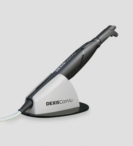
Our CariVu camera uses safe, radiation-free light transillumination to detect cavities and cracks that can be difficult to see with the naked eye or on a traditional X-ray. Light passes through the tooth, and areas of decay appear as shadows, allowing our team to identify problems at their earliest stages.
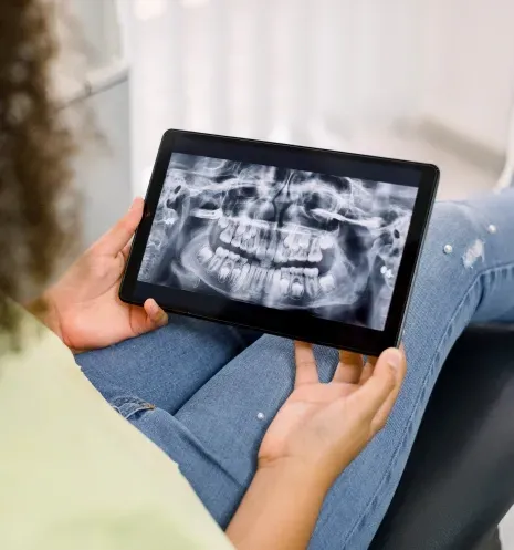
Digital X-rays allow us to capture clear, detailed images of your teeth using significantly less radiation than traditional film X-rays. Because the images appear instantly on screen, our team can review them with you in real time and explain findings as they come up.
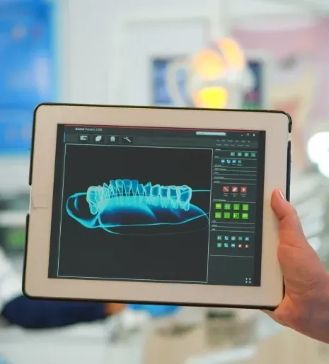
We use AI supported diagnostic software to help review X-rays and scans with an added layer of precision. The technology flags areas of potential concern, such as early decay or bone loss, so our team can evaluate them more closely alongside their own clinical judgment.
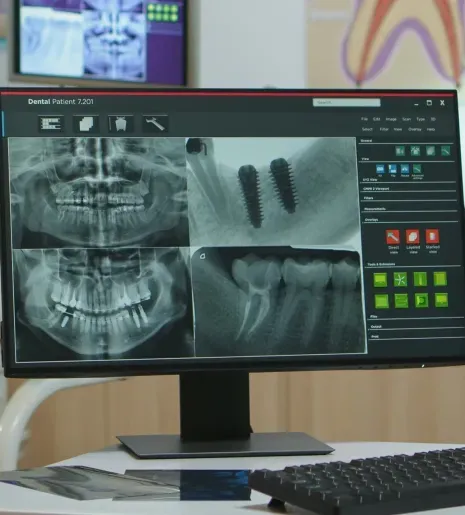
For patients undergoing full mouth implant treatment, we use a fully digital workflow that maps out the entire procedure before treatment begins. This allows our team to plan implant placement with precision, anticipate challenges in advance, and design a smoother path from start to finish.
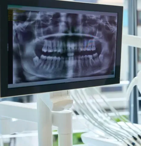
Once implants are placed, we use digital design tools to create custom restorations that are built to match your bite, function, and natural smile. This digital approach allows for a more precise fit and a more efficient path to your final restoration.
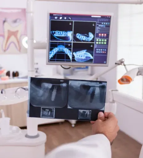
We partner with a high end dental laboratory to fabricate restorations with an exceptional level of craftsmanship and detail. This partnership allows us to combine our in-house digital technology with skilled lab work for results that look natural and fit precisely.
Common Questions
In most cases, yes. Our intra oral scanners capture highly accurate 3D digital impressions of your teeth and bite in just a few minutes, eliminating the need for the thick, uncomfortable putty trays used with traditional impressions. These digital scans are used for crowns, aligners, implant planning, and more, and they also allow you to see a digital model of your own teeth on screen during your visit.
Digital X-rays use significantly less radiation than traditional film X-rays while producing instant, high-resolution images. Because the images appear on screen right away, our team can review them with you in real time, explain what we are seeing, and easily compare images over time to track changes in your oral health, all without the wait time associated with developing traditional film.
No. Our AI supported diagnostic software works alongside, not in place of, our dentists' clinical expertise. It helps identify early signs of decay, bone loss, and other concerns, and adds a second layer of review to support our team's diagnosis. The findings are highlighted visually, which makes it easier for our team to explain what they see and for you to understand your treatment recommendations.
CariVu uses safe, radiation-free light transillumination rather than radiation to detect cavities and cracks. Light passes through the tooth, and areas of decay appear as shadows, which allows our team to identify problems at their earliest stages, including cavities between teeth and cracks that are not always visible on a traditional X-ray. Because it uses light instead of radiation, it is safe to use as often as needed.
For patients undergoing full mouth implant treatment, we use a fully digital workflow that maps out implant placement using detailed 3D imaging and digital scans before treatment begins. This reduces guesswork and surprises during surgery, supports better coordination between planning, placement, and final restoration, and helps set realistic expectations for your treatment timeline.
Yes. Our intra oral cameras and scanners display images on a chairside monitor in real time, so you can see exactly what your dentist sees. Digital X-rays appear instantly on screen as well, allowing our team to review findings with you as they come up, making exams more transparent and easier to understand.
Real Results
See how implants, whitening, and same-day crowns have restored confidence for patients in Burtonsville. Results vary; we will show you what is realistic for your smile during a consultation.
See More Smiles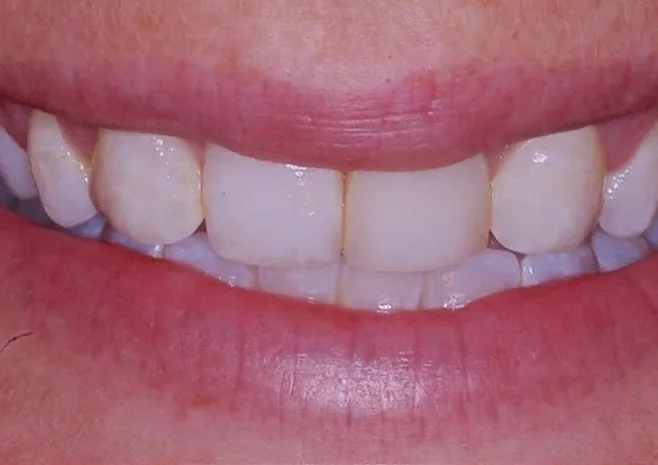 Before
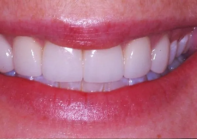 After
After
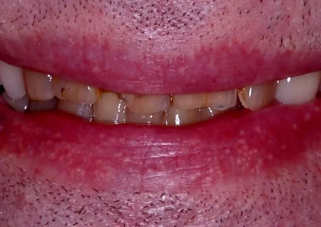 Before
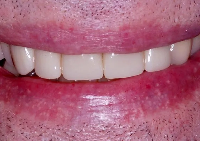 After
After
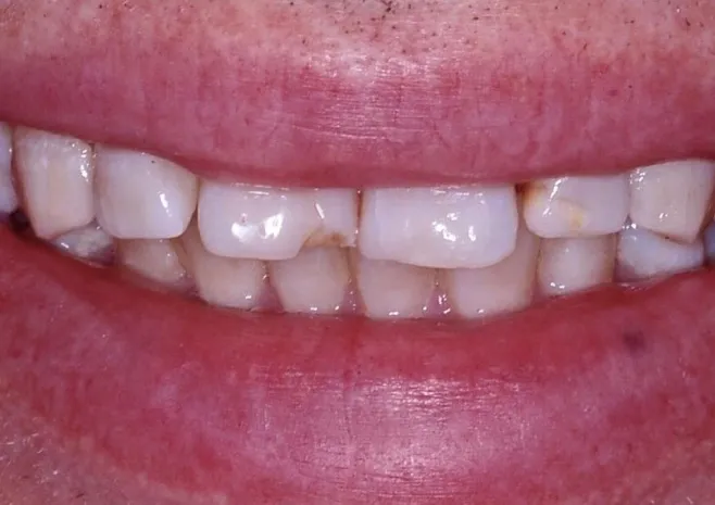 Before
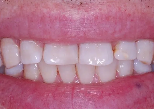 After
After
 4009 Sandy Spring Road, Suite 201, Burtonsville, MD 20866

 301-622-1717